Which Part of the Neuron Can Conduct an Action Potential
Cardinal facts: action potential and synapses
- Neurons communicate with each other via electric events called 'activity potentials' and chemic neurotransmitters.
- At the junction between two neurons (synapse), an activeness potential causes neuron A to release a chemical neurotransmitter.
- The neurotransmitter tin either help (excite) or hinder (inhibit) neuron B from firing its own action potential.
- In an intact encephalon, the remainder of hundreds of excitatory and inhibitory inputs to a neuron determines whether an activeness potential volition result.
Neurons are essentially electrical devices. In that location are many channels sitting in the cell membrane (the purlieus betwixt a cell'south inside and exterior) that let positive or negative ions to flow into and out of the jail cell.
Usually, the inside of the prison cell is more negative than the exterior; neuroscientists say that the inside is around -70 mV with respect to the exterior, or that the cell'due south restingmembrane potential is -70 mV.
This membrane potential isn't static. It'due south constantly going up and downwards, depending mostly on the inputs coming from the axons of other neurons. Some inputs make the neuron's membrane potential become more than positive (or less negative, eastward.g. from -seventy mV to -65 mV), and others do the opposite.
These are respectively termed excitatory and inhibitory inputs, as they promote or inhibit the generation ofaction potentials (the reason some inputs are excitatory and others inhibitory is that different types of neuron release different neurotransmitters; the neurotransmitter used by a neuron determines its upshot).
Action potentials are the primal units of communication between neurons and occur when the sum total of all of the excitatory and inhibitory inputs makes the neuron's membrane potential attain around -50 mV (see diagram), a value chosen theaction potential threshold.
Neuroscientists oftentimes refer to action potentials every bit 'spikes', or say a neuron has 'fired a fasten' or 'spiked'. The term is a reference to the shape of an activity potential as recorded using sensitive electrical equipment.
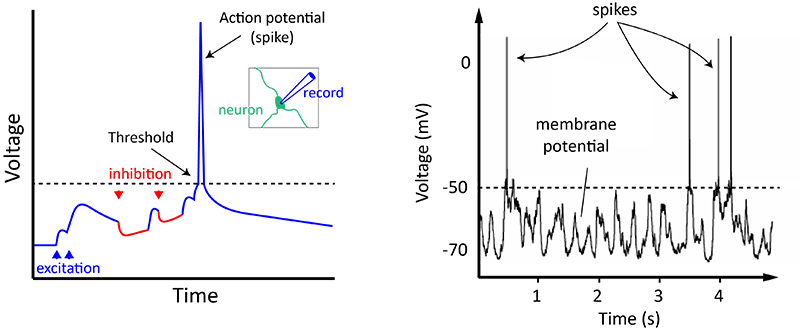
A neuron spikes when a combination of all the excitation and inhibition it receives makes it accomplish threshold. On the right is an example from an actual neuron in the mouse's cortex. (Epitome: Alan Woodruff / QBI)
Synapses: how neurons communicate with each other
Neurons talk to each other acrosssynapses. When an action potential reaches the presynaptic terminal, it causes neurotransmitter to be released from the neuron into thesynaptic cleft, a xx–40nm gap between thepresynaptic axon last and thepostal servicesynaptic dendrite (oftentimes a spine).
After travelling across the synaptic scissure, the transmitter will adhere to neurotransmitter receptors on the postsynaptic side, and depending on the neurotransmitter released (which is dependent on the type of neuron releasing it), particular positive (due east.g. Na+, Yard+, Ca+) or negative ions (e.g. Cl-) will travel through channels that bridge the membrane.
Synapses can be thought of as converting an electrical signal (the activeness potential) into a chemical point in the course of neurotransmitter release, and then, upon binding of the transmitter to the postsynaptic receptor, switching the indicate back again into an electrical form, as charged ions period into or out of the postsynaptic neuron.
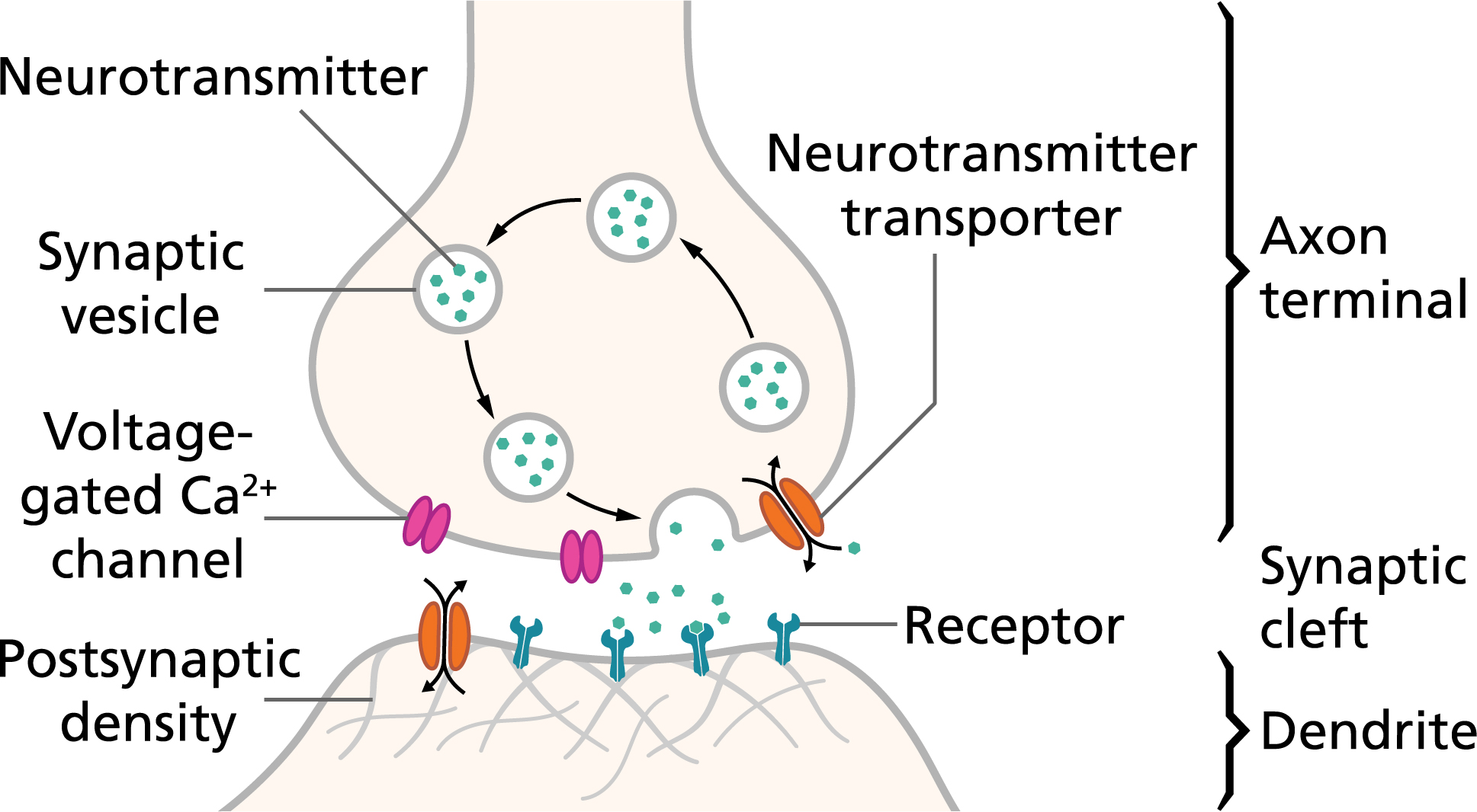
An action potential, or spike, causes neurotransmitters to be released across the synaptic scissure, causing an electrical signal in the postsynaptic neuron. (Image: By Thomas Splettstoesser / CC BY-SA 4.0)
Video: Action potentials in neurons
Concepts and definitions
Axon – The long, thin structure in which activity potentials are generated; the transmitting part of the neuron. After initiation, activity potentials travel downward axons to crusade release of neurotransmitter.
Dendrite – The receiving part of the neuron. Dendrites receive synaptic inputs from axons, with the sum full of dendritic inputs determining whether the neuron will fire an action potential.
Spine – The small protrusions institute on dendrites that are, for many synapses, the postsynaptic contact site.
Membrane potential – The electrical potential across the neuron's jail cell membrane, which arises due to unlike distributions of positively and negatively charged ions within and outside of the cell. The value within of the cell is always stated relative to the exterior: -lxx mV means the inside is 70 mV more negative than the outside (which is given a value of 0 mV).
Action potential– Brief (~1 ms) electrical issue typically generated in the axon that signals the neuron as 'agile'. An activity potential travels the length of the axon and causes release of neurotransmitter into the synapse. The action potential and consistent transmitter release allow the neuron to communicate with other neurons.
Neurotransmitter –A chemical released from a neuron following an action potential. The neurotransmitter travels across the synapse to excite or inhibit the target neuron. Dissimilar types of neurons use dissimilar neurotransmitters and therefore have different furnishings on their targets.
Synapse – The junction between the axon of ane neuron and the dendrite of another, through which the two neurons communicate.
QBI research
QBI Laboratories working on neurons and neuronal communication: Professor Stephen Williams, Professor Pankaj Sah
QBI Laboratories working on synapses: Dr Victor Anggono, Professor Joseph Lynch, Professor Frederic Meunier
traviswitteorsell.blogspot.com
Source: https://qbi.uq.edu.au/brain-basics/brain/brain-physiology/action-potentials-and-synapses
Belum ada Komentar untuk "Which Part of the Neuron Can Conduct an Action Potential"
Posting Komentar